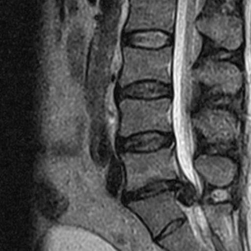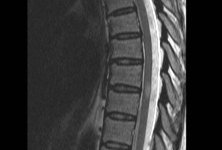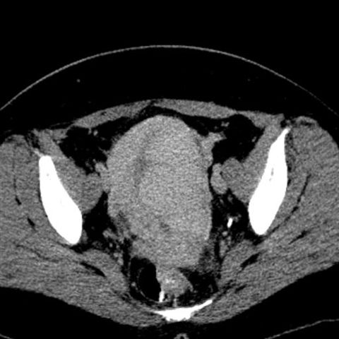
Body Imaging (Abdomen & Pelvis)
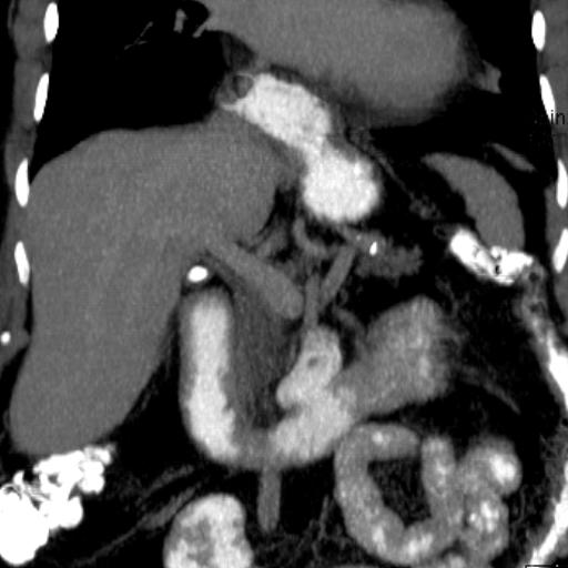
The abdomen and pelvis are usually studied initially by sonography because it is less expensive, widely available, and does not use ionizing radiation. Subsequently and depending on the particular clinical application, CT scans are obtained when it is necessary to clarify sonogram findings.