Nuclear Medicine
SPECT-CT studies should be requested by the referring physician.
Pulmonary Studies
Endocrine Studies
Neuro / Brain Studies
Musculoskeletal Studies
Infections or Tumor Studies
Gastrointestinal Studies
Renal Studies
Cardiac Studies
All Nuclear Medicine Studies
To reduce radiation exposure, patients should be well hydrated prior to and for at least one day after injection.
Gastrointestinal Studies
Preparation: Your last meal should be no less than four hours before the study.
Duration: Minimum of two hours.
Indications: acute cholecystitis, chronic cholecystitis, bile duct obstruction, biliary extravasation, biliary atresia, liver transplant evaluation, vague gallbladder (ejection fraction calculation) .
Gastric Emptying is a study where the emptying of the stomach is evaluated. It is a study where the patient eats the egg white stir fry mixed with the radioactive material and images are acquired at 1 minute, 15 minutes, 1hr, 2hr, and 4hr (optional).
Preparation: Your last meal should be no less than four hours before the study. Should bring three raw eggs, bread, jam and juice.
Duration: Minimum of five hours.
Pulmonary Studies
Preparation: none required.
Duration: 30 to 45 minutes.
Indications: regional ventilation evaluation.
Preparation: none required.
Duration: 30 to 45 minutes.
Indications: possible PE.
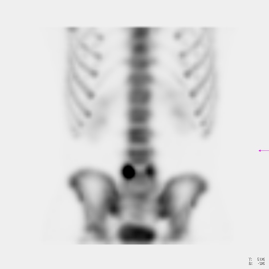
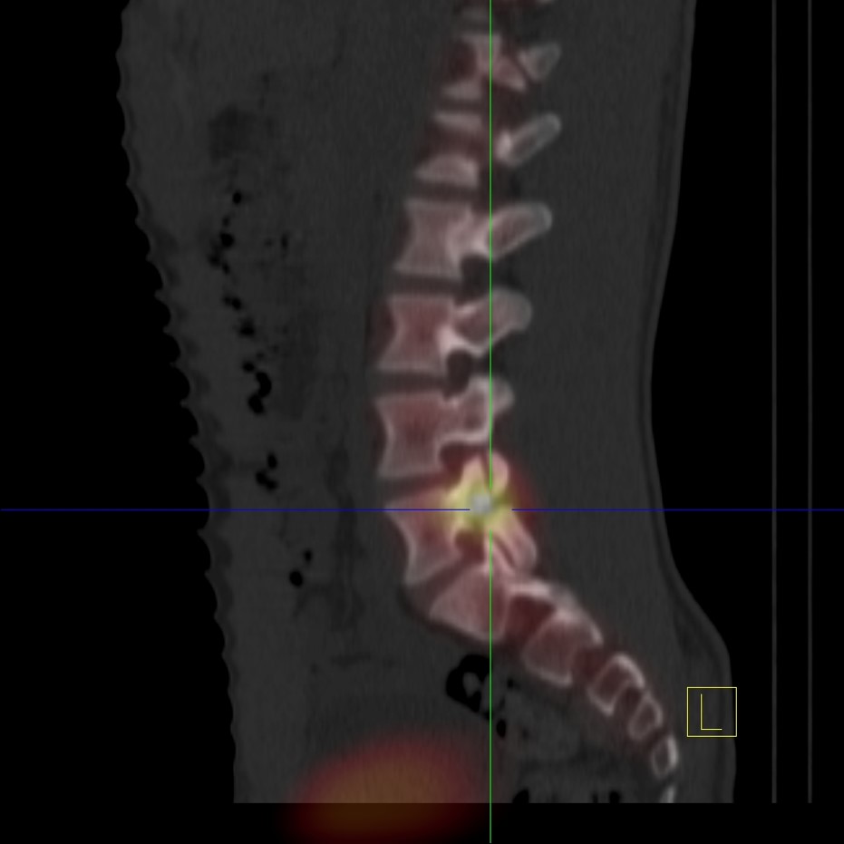
Musculoskeletal Studies
Preparation: none required.
Duration 45 minutes to one hour.
Indications: arthritis, primary tumors, metastases, fractures not identified or equivocal in radiology studies.
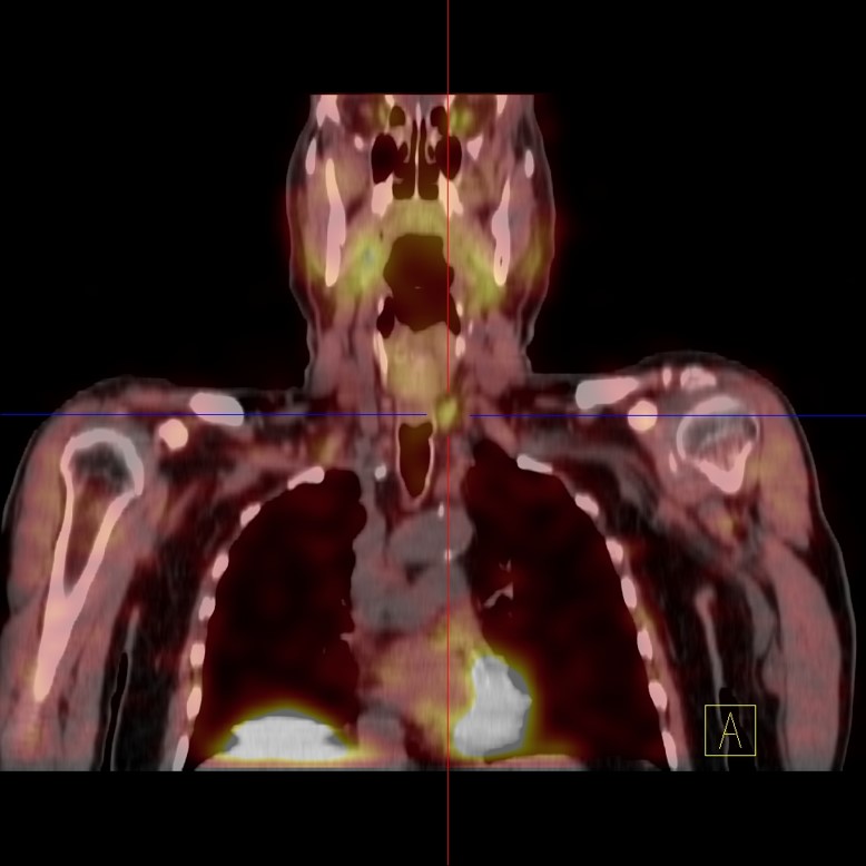
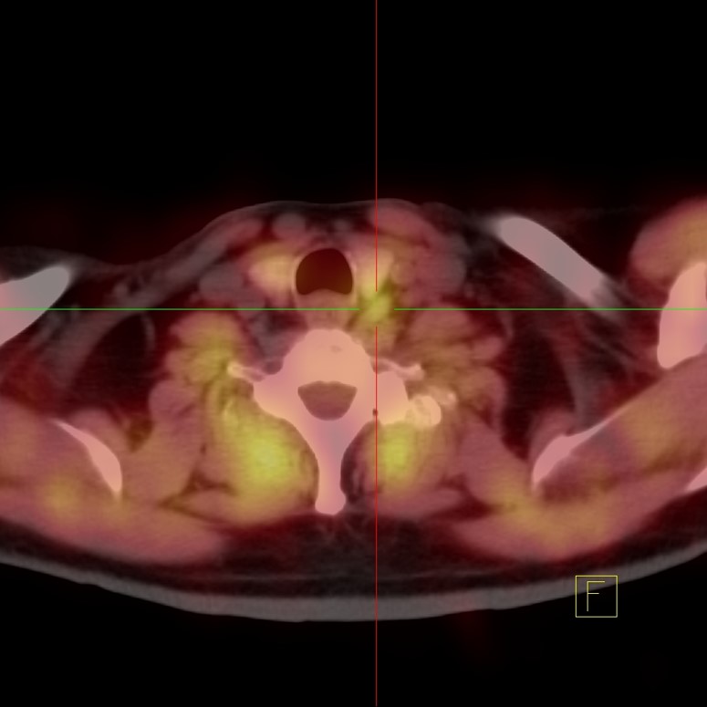
Infections or Tumors Studies
Preparation: none required.
Duration: one hour.
Indications: infections, inflammation, or tumors.
Renal Studies
Preparation: Hydration prior to and during the study.
Duration: one hour to one and a half hour.
Indication: kidney failure, renal hypertension, kidney transplant, renal obstruction, and tubular function and secretion.
Cardiac Studies
Preparation: none required.
Duration: one hour.
Indications: Quantify ventricular function.
Preparation: Your last meal should be no less than four hours before the study.
Duration: one hour.
Indications: Infarct within the heart walls.
MEDFLIX performs a CT coronary calcium score study at no additional cost to MPI patients who do not have coronary stents and who have not undergone coronary artery bypass grafting. This improves the specificity of MPI findings.
Preparation: Your last meal should be no less than four hours before the study. No coffee, chocolate, tea, or soda drinks 12 hours prior to the study.
Duration: half day.
Indications: Possible cardiac ischemia or infarction.
Neuro/Brain Studies
Preparation: none
Duration: one hour
Indications: neurodegenerative dementia and epilepsy
Preparation: Discontinue medications, if applicable.
Duration: six to eight hours.
Indications: To differentiate between essential tremors and parkinsonian syndromes.
Endocrine Studies
Preparation: A week prior to the study, stop taking all thyroid medications.
Duration: 30 minutes.
Indications: hyperthyroidism, hypothyroidism, detect nodules, chronic thyroiditis, Hashimoto, hyperfunctional adenoma, thyroid cancer, and evaluation of ectopic thyroid tissue
The Parathyroid Scan with SPECT-CT is for the localization of parathyroid adenomas in the setting of overactive parathyroid glands.
Preparation: none.
Duration: three hours.
Indications: Detect parathyroid adenomas.
The images obtained with I-131 in a SPECT-CT scanner provide significantly more information than conventional planar studies, including those using pinhole collimation, thanks to the anatomic localization studies and precision that a CT image offers when superimposed in the traditional nuclear medicine image.
Preparation and Duration: Depends if the I-131 is used for treatment or to perform a scan. For details please contact our Communications Center.
Indications: hyperthyroidism and cancer therapy
Preparation: The use of laxatives should be considered when the abdomen is the area of interest. A mild oral laxative may be administered the evening prior to injection and the evening following injection. The need for bowel preparation should be assessed on an individual basis.
Duration: Whole body images are obtained at four and 24 hours after tracer injection. SPECT-CT images are obtained at 24 hours.
2. Staging patients with neuroendocrine tumors.
3. Follow-up of patients with the known disease to evaluate potential recurrence.
Weight Limit
500 Pounds. The weight must be corroborated at MEDFLIX with a weight scale that registers up to 500 lbs the day of the study.