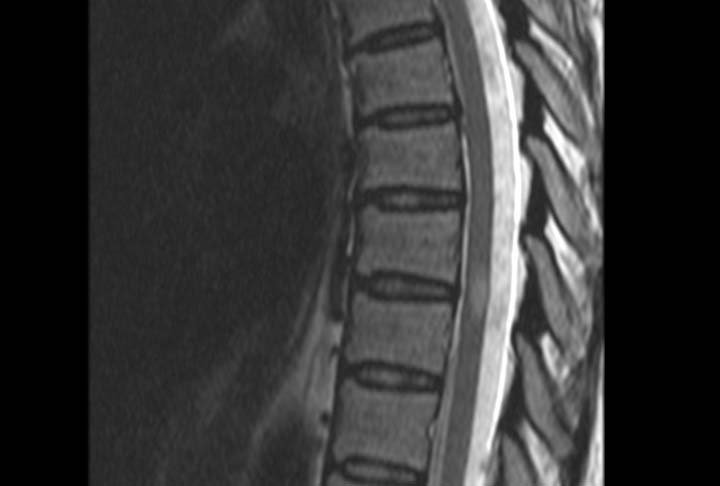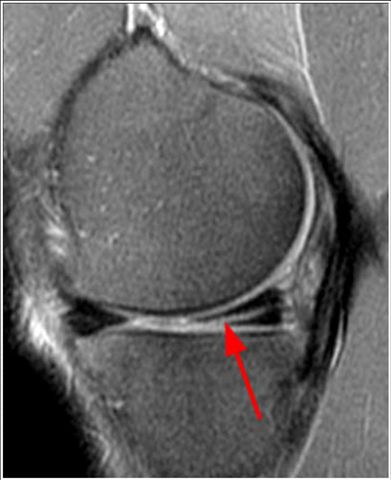
Image 1
MRI of the knee, sagittal/lateral magnified image to the medial meniscus (interior). The red arrow shows a tear of the posterior horn of the meniscus visualized by a bright line. The healthy meniscus looks uniformly black
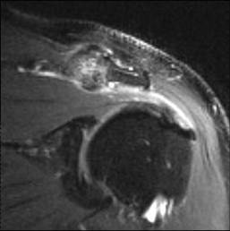
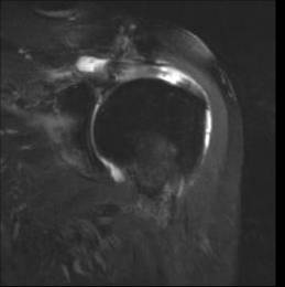
Image 2
MRI shoulder, coronal/frontal image. The joint between the clavicle and acromial process of the scapula demonstrates severe arthritic changes that compress the muscle and main tendon of the rotator cuff, the supraspinatus. The healthy tendon should appear black. The tendon looks shiny and irregular rather than black indicating tendonitis. The bursa located between the acromioclavicular and supraspinatus joints and the deltoid is thickened by bright fluid indicating bursitis
