Breast MRI does not replace mammography, but complements it. All breast MRI studies should be interpreted and correlated with a recent mammogram and often with a recent sonomammography. Any breast MRI study intended to detect breast cancer or other pathology requires the injection of intravenous gadolinium contrast. Both breasts should be studied routinely since this modality detects cancer that is not clinically suspected in other breast studies in the breast as opposed to the one that is suspected or has already been documented to have a problem. It also detects cancer in other areas of the breast where cancer is suspected or already documented. This information may change the type of treatment to be followed. If there has been a mastectomy, total or partial, residual tissue, including the anterior chest wall, should be included.
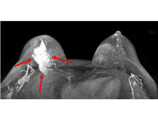
Image 1
Large red arrowed mass in the right breast represents primary cancer
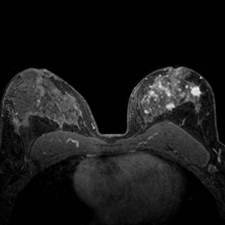
Image 2
Multiple small masses in the left breast represent much more extensive multicentric cancer than this patient's mammography suggested. The opposite breast also showed a small cancer invisible by mammography. Both breasts should be studied routinely, not just the breast of concern
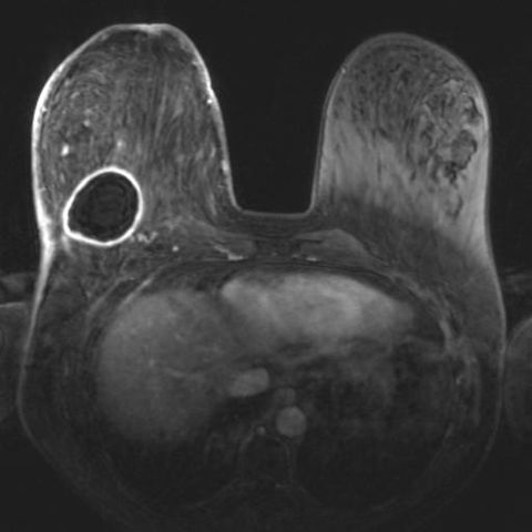
Image 3
Ring-shaped, contrast-colored lesion on the back of the right breast represents an abscess (infection). Not all breast lumps are cancerous
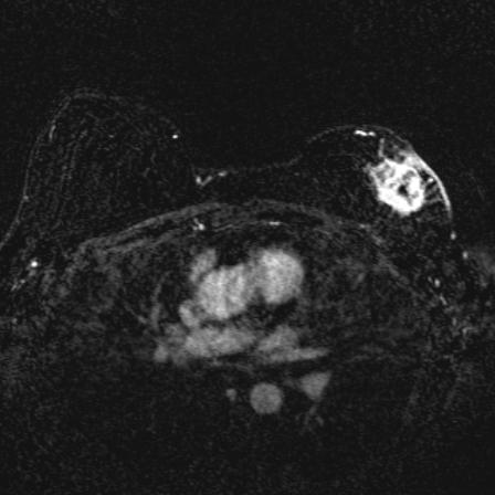
Image 4
Patient had left breast cancer treated with lumpectomy and radiation presents with palpable mass near surgical scar. Breast MRI shows an irregular lesion that captures contrast in the front and outside of the left breast extending to the skin. Biopsy was positive for local recurrence
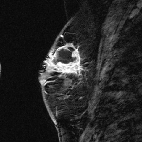
Image 5
Same patient image 4 demonstrated the extent of this recurrent cancer unfolded from another angle, sagittal or lateral perspective
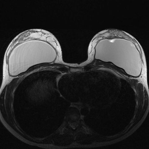
Image 6
Intact breast implants with no evidence of rupture MRI is the modality par excellence for the detection of intracapsular and extracapsular breast implant ruptures
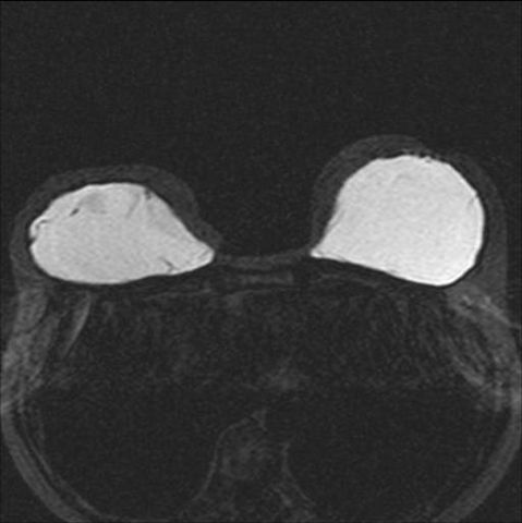
Image 7
Breast implants with evidence of intracapsular rupture The black wavy lines recall the appearance of the linguine paste strips as they are formally named when reported in radiology reports
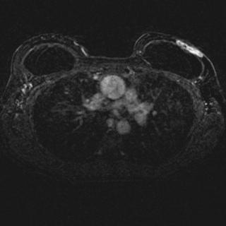
Image 8
Breast implant patient with breast cancer in front of his implant. Mammography in patients with breast implants does not show all glandular tissue due to the presence of the implant. Implants also preclude optimal compression during mammography
Would you like to schedule an appointment?
Now you can make appointments online with San Patricio MEDFLIX.