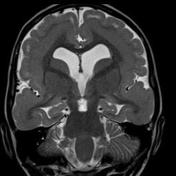
- Brain
- Orbits
- Paranasal Sinuses / Maxillofacial
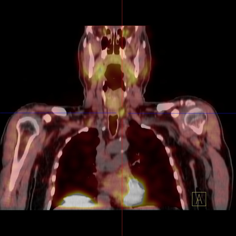
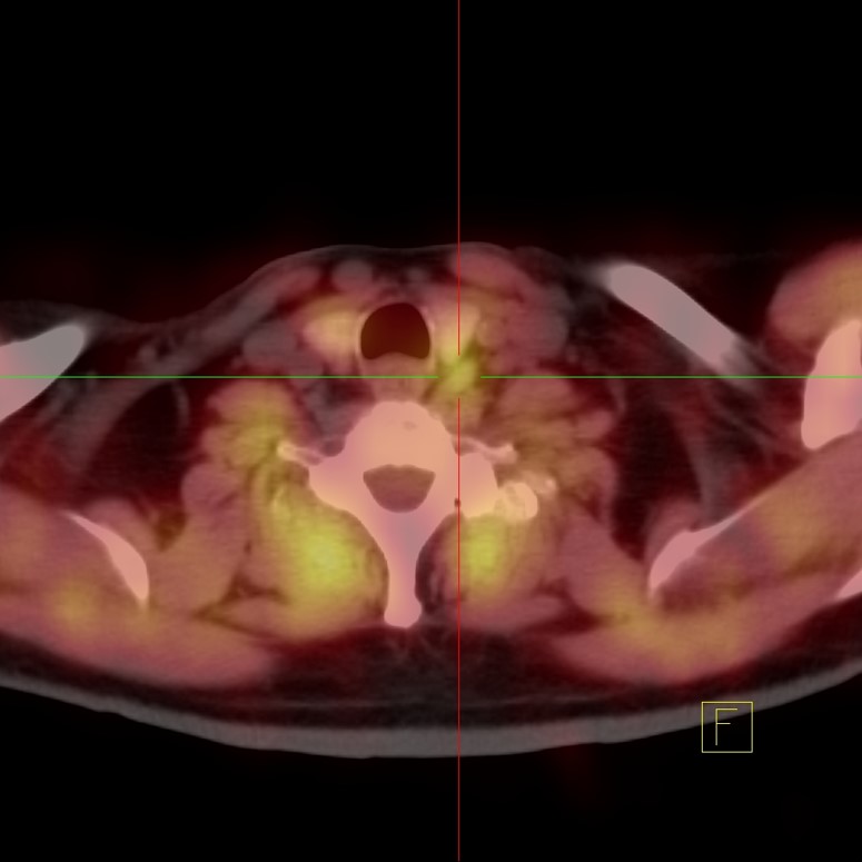
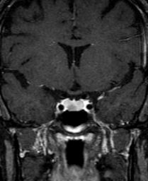
- Neck
- Spinal Column & Spinal Cord
- Cervical
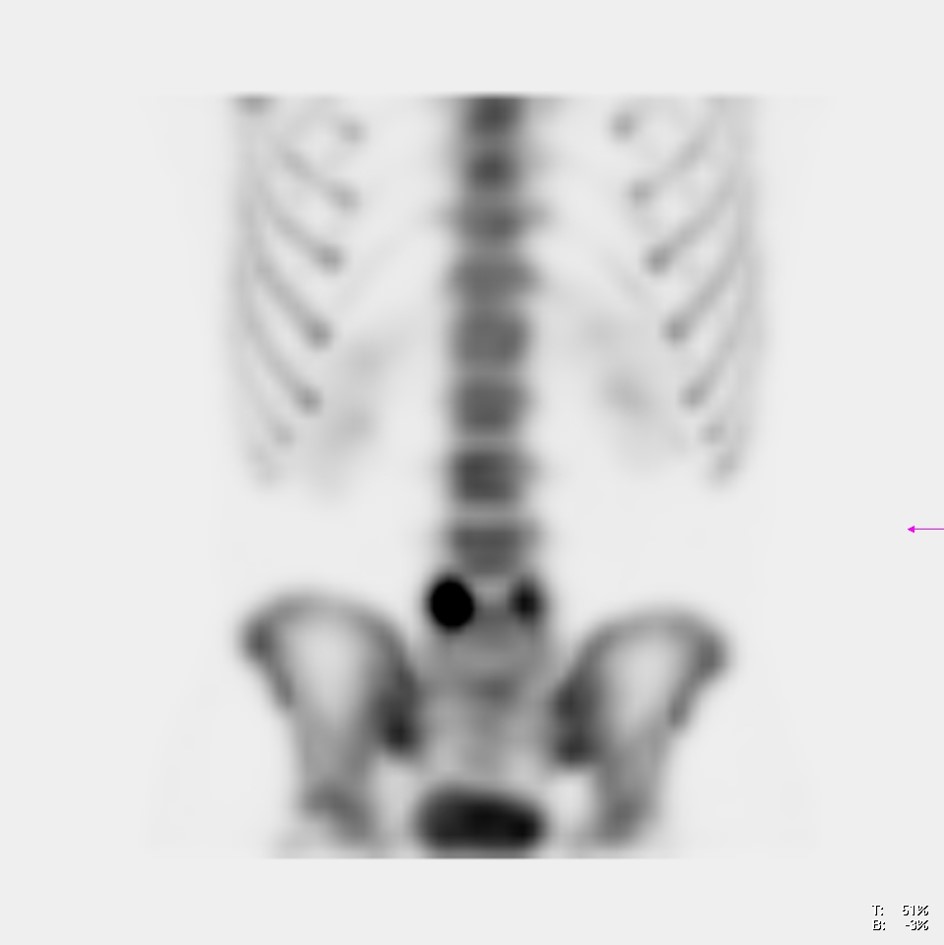
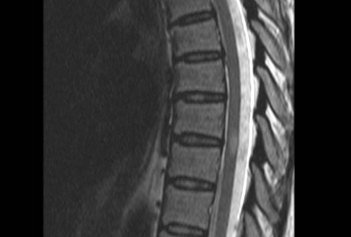
- Thoracic / Dorsal
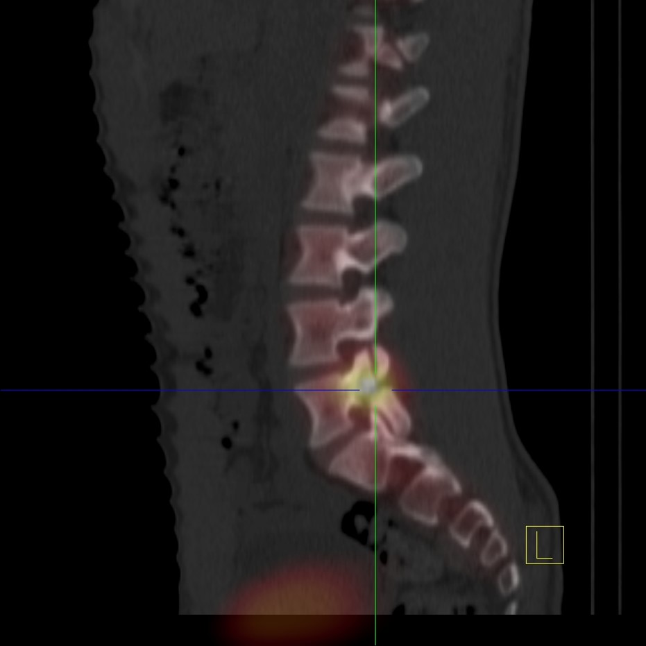
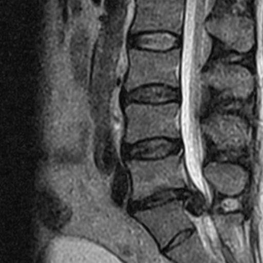
- Lumbar
- Sacral
- Sacroiliac Joints
- TMJ (Temporomandibular joint)
- Prostate – Multiparametric Protocol
- Scrotum/Testicles
- Anorectal
Musculoskeletal Studies
- Upper Extremities (without and with IV
contrast when considering arthropathies)- Shoulder
- Forearm
- Elbow
- Wrist
- Hand
Musculoskeletal Studies
- Lower Extremities (without and with IV contrast
when considering arthropathies)- Hip
- Thigh
- Calf
- Ankle
- Foot
- Endometrium
- Vagina
- Anorectal
Body Studies
- Chest
- Breasts
Body Studies
- MRCP (Magnetic Resonance Cholangiopancreatography)
- Abdomen
- Liver
- Pancreas
- Adrenals
- Pelvis
- Brain
- Neck
- Spinal Column and Spinal Cord
- Cervical
- Thoracic/Dorsal
- Lumbar
- Sacral
- Sacroiliac Joints
- Musculoskeletal
- Chest
- Abdomen
- Liver
- Pancreas
- Adrenals
- Pelvis
- Lower Extremities