Musculoskeletal

Share on facebook
Share on twitter
Share on pinterest

Image 1

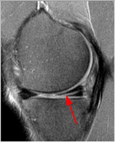
MRI of the knee, sagittal/lateral magnified image to the medial meniscus (interior). The red arrow shows a tear of the posterior horn of the meniscus visualized by a bright line. The healthy meniscus looks uniformly black

Image 2

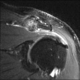
MRI shoulder, coronal/frontal image. The joint between the clavicle and acromial process of the scapula demonstrates severe arthritic changes that compress the muscle and main tendon of the rotator cuff, the supraspinatus. The healthy tendon should appear black. The tendon looks shiny and irregular rather than black indicating tendonitis. The bursa located between the acromioclavicular and supraspinatus joints and the deltoid is thickened by bright fluid indicating bursitis

Image 3

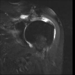
MRI shoulder, coronal/frontal image. The supraspinous tendon demonstrates a focal interruption delineated by bright fluid communicating the bursa with the humeral joint and scapula (glenohumeral). Complete rupture of the tendon

Would you like to schedule an appointment?

Now you can make appointments online with
San Patricio MEDFLIX.

Subscribe to our Newsletter

Access information about a variety of interesting topics about the world of radiology and nuclear medicine medical imaging.

Share this post with your friends

Share on facebook
Share on twitter
Share on linkedin

Hours of Operation

Monday - Thursday

6:00am - 4:00pm

Phone Hours

Monday - Friday

6:00am - 4:00pm

Contact Information

787-620-5761

[3cx-clicktotalk id="626" title="Live Chat & Talk item 1"]

Copyright 2020-2021 - San Patricio MEDFLIX. All rights reserved. Made tasty by Mango Media.

Parathyroid scan, SPECT-CT, fused image - parathyroid adenoma below left thyroid lobe

Parathyroid scan, SPECT-CT, fused image - parathyroid adenoma behind left lobe of thyroid

Bone scan SPECT - bilateral posterior L5 uptake

Bone scan SPECT-CT - fused sagittal image confirms L5 spondylolysis

CT Chest - Bronchioloalveolar Cell Carcinoma

CT Abdomen Esophageal Hernia

CT Parotid Glands - Sjogren's syndrome - sialolithiasis

CT 3D Lumbar Hardware

Brain MRI, pediatric - Lissencephaly and band heterotopia

Brain MRI, pituitary, elevated prolactin - prolactinoma

Lumbar spine MRI - extruded L5-S1 disk

Thoracic spinal cord MRI - multiple sclerosis