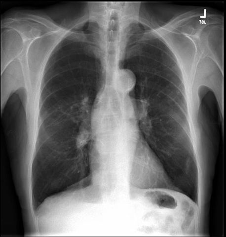
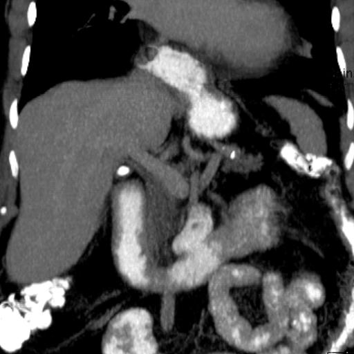
Chest CT with intravenous contrast demonstrates aneurysm of the ascending thoracic aorta correlating with imaging x-rays 1 and 2
Image 4
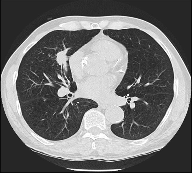
Chest CT with intravenous contrast dedicated to the pulmonary arteries demonstrates a filling defect in the left pulmonary artery. This is the typical appearance of a pulmonary embolism. It is mandatory to inject contrast to make this CT diagnosis. The diagnosis is not made with chest x-rays although these are obtained as an initial evaluation
Image 5
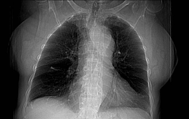
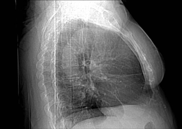
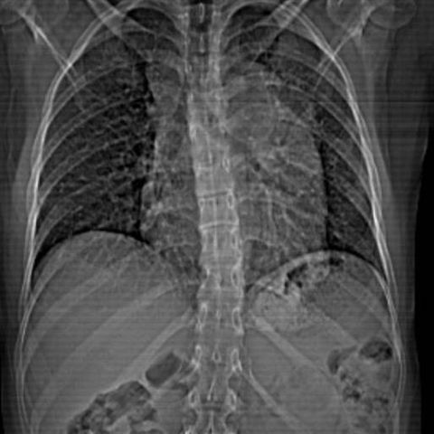
Frontal chest x-ray shows abnormally enlarged mediastinum. Chest CT without and with intravenous contrast is indicated to evaluate what produces the X-ray finding
Image 6
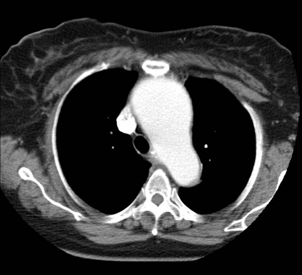
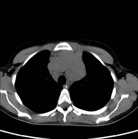
Chest CT without intravenous contrast confirms that there is a mass in the mediastinum excluding aortic aneurysm. The non-contrast intravenous image also rules out calcifications in the mass
Image 7
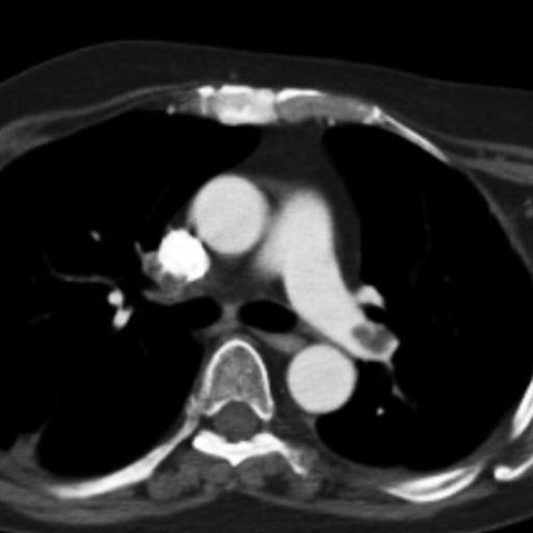
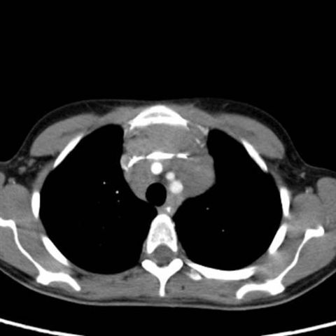
Chest CT with intravenous contrast shows that the mass compresses the main arteries in the mediastinum. The mass turned out to be Hodgkin's lymphoma
Image 8
Frontal chest x-ray shows emphysema without evidence of mass
Image 9
Breast CT shows a non-calcified mass of concern for cancer in the right lung, invisible on x-ray
Image 10
Image 11
Image 12
Image 13
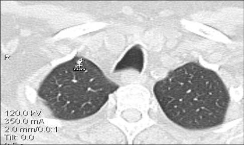
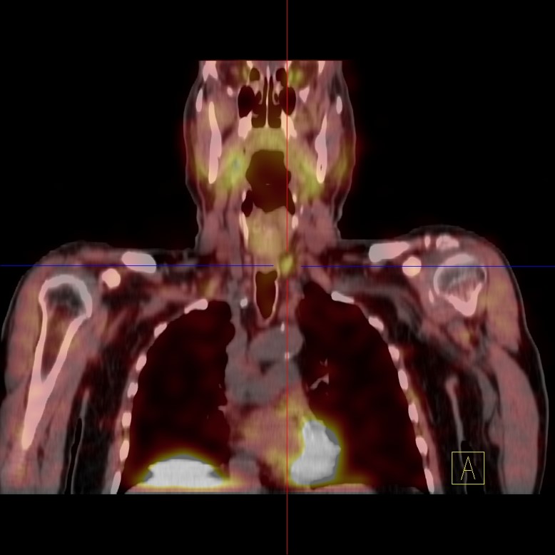
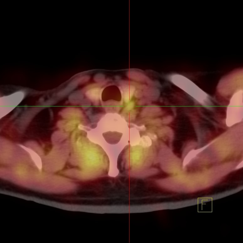
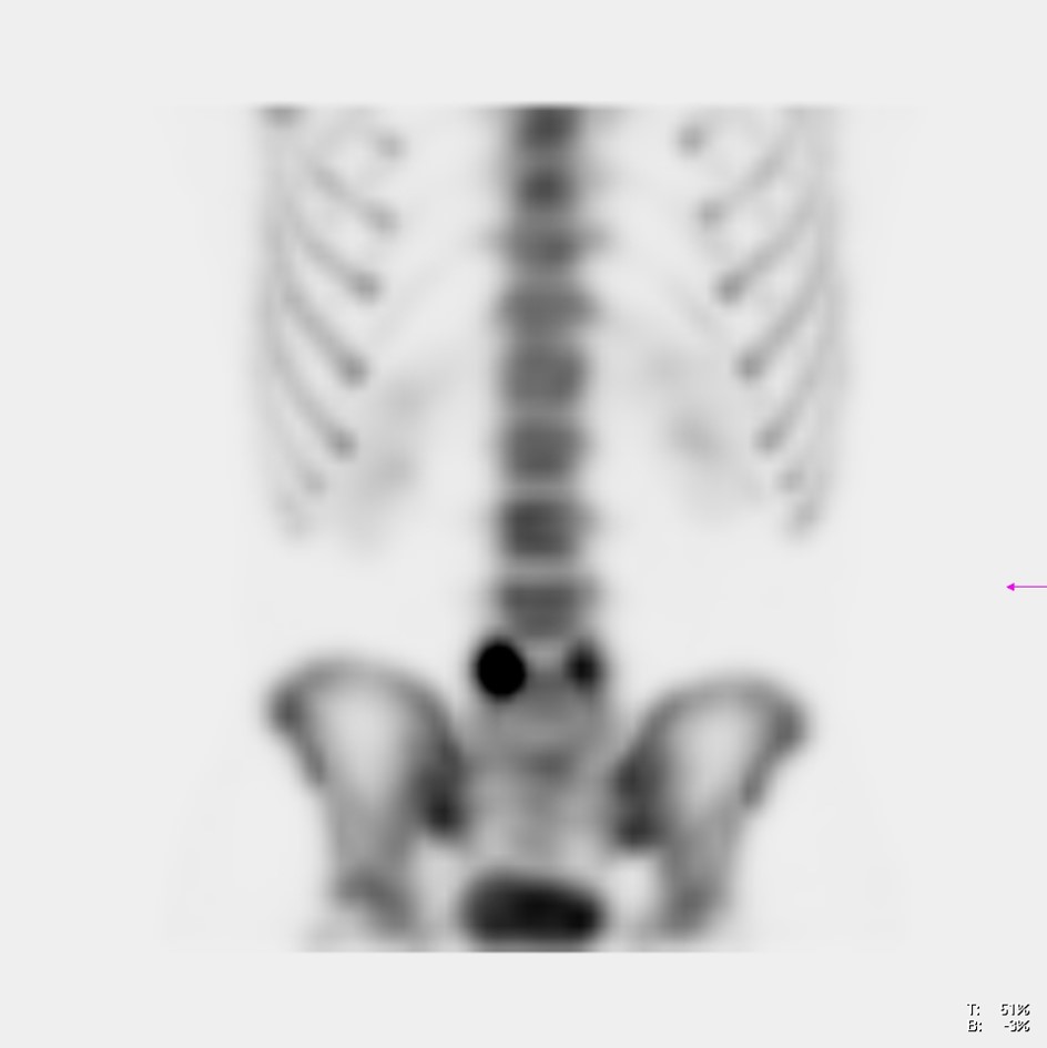
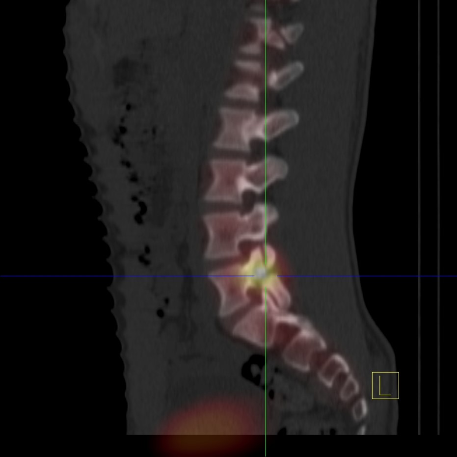
Serial chest CT scans obtained every three months demonstrating the progressive growth of a lung nodule from less than one centimeter to a cancerous mass originating in the right lung and invading the pleura, confirmed by biopsy. From the nodule demonstrating unresponsive growth, a biopsy should be performed as soon as possible to document cancer and determine its cell type. A PET-CT scan from the head to the hips is needed to determine how extensive the disease is (staging)
Image 14
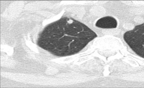
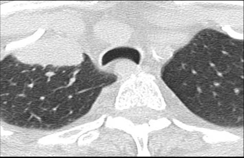
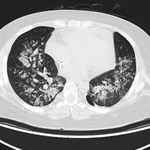
Non-contrast intravenous chest CT demonstrates an irregular, non-calcified nodule in contact with the posterior pleura of the right lung
Image 15
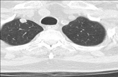
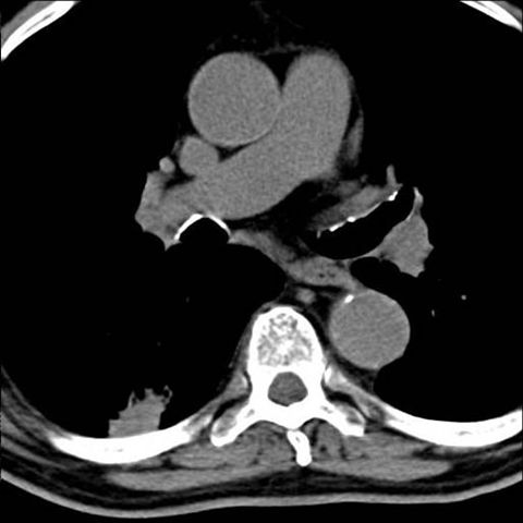
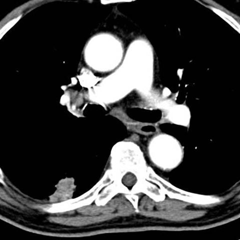
CT of the chest with intravenous contrast shows that the node is enhanced with contrast and that the right floss has a prominent lymph node that is invisible in the image without intravenous contrast. Findings represent a right lung cancer and metastasis to the right floss. A PET-CT scan from head to hips is needed to determine how extensive the disease is (staging)
Would you like to schedule an appointment?
Now you can make appointments online with San Patricio MEDFLIX.